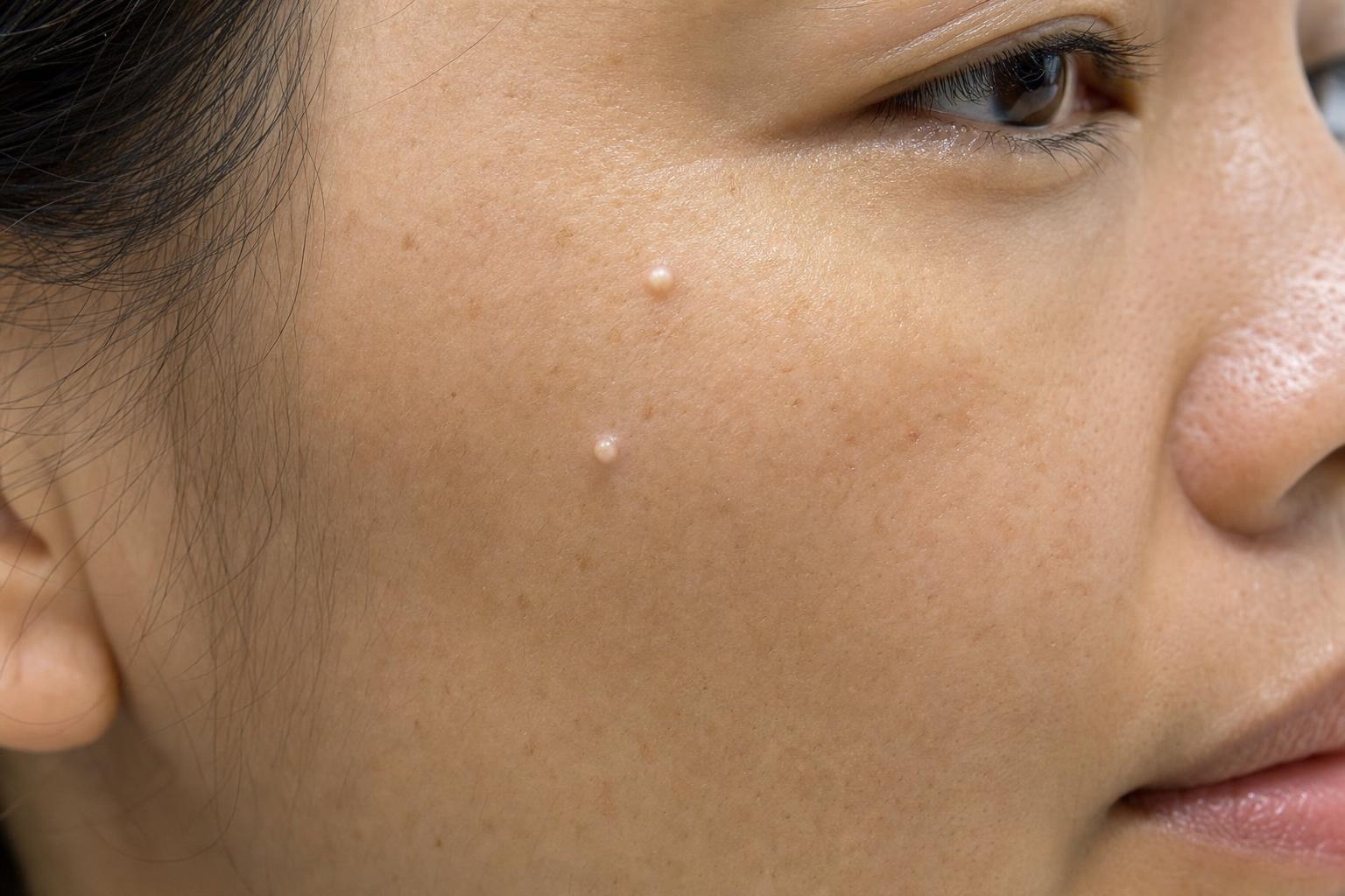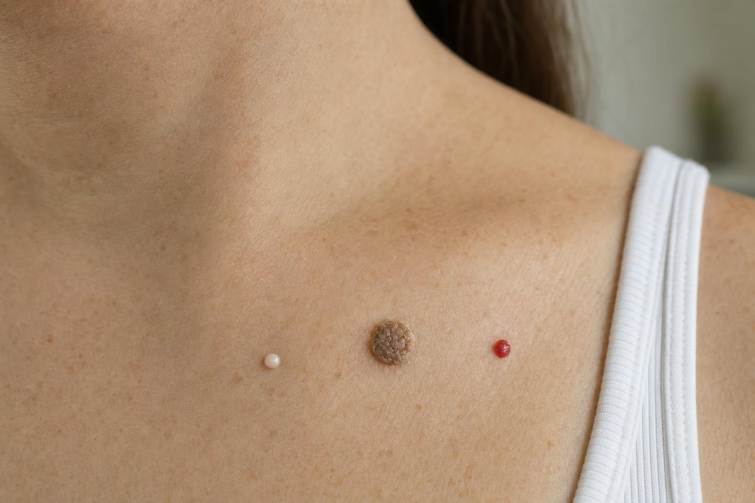
A seborrheic keratosis often looks waxy, rough or as though it has been pasted onto the skin. A mole is formed by pigment-producing cells and is more likely to have a smooth, consistently coloured surface. Those are useful clues—not a diagnosis.
Both growths can be flat or raised, tan or nearly black, and regular or irregular. Melanoma and other skin cancers can also resemble harmless spots. The practical rule is simple: if a growth is new, changing, unusual for you, repeatedly bleeding or otherwise uncertain, arrange a medical assessment before considering cosmetic removal.
This distinction matters because seborrheic keratoses are benign, and most moles are harmless, but a suspicious lesion should not be treated as a cosmetic problem until its identity is clear.
Key takeaways
- A waxy, scaly or “stuck-on” surface commonly points toward a seborrheic keratosis, but it is not definitive.
- Typical moles are often symmetrical, evenly coloured and stable over time, although harmless moles vary considerably.
- Evolution is one of the most important warning signs: watch for changes in size, shape, colour, surface or symptoms.
- A lesion that looks different from your other spots deserves attention even if it does not meet every standard warning sign.
- Professional removal is appropriate only after a growth is understood to be benign and suitable for cosmetic treatment.
How these growths usually differ
Seborrheic keratoses and moles arise from different types of cells, but that biological difference is not always obvious at home. It is more useful to compare their usual texture, outline, colour and history.
Typical features of a seborrheic keratosis
A seborrheic keratosis is a non-cancerous growth arising from the outer layers of the skin. These growths become more common with age and may appear on the torso, face, scalp, shoulders or other areas. They do not usually develop on the palms or soles.
Common features include:
- A waxy, rough, scaly or slightly crumbly surface
- A raised appearance that can look pasted or stuck onto the skin
- Colour ranging from pale tan to brown or black
- A round or oval shape, sometimes with an uneven outline
- One isolated growth or many similar growths
Some are thin and only slightly raised. Others become thick, textured and easy to irritate with a collar, bra strap, waistband or jewellery. A dark or inflamed keratosis can look particularly concerning, so texture alone should not be used to dismiss it.
Typical features of a mole
A mole, also called a nevus, is a collection of melanocytes—the cells that produce pigment. Moles may be present from childhood or develop later. A typical benign mole is often round or oval, symmetrical and stable, with one consistent shade of tan, brown or another colour.
Moles can be flat, dome-shaped or raised. Their surfaces may be smooth or slightly textured, and hair may grow through them. Normal moles do not all look identical, especially when comparing different people or skin tones. What matters is whether a particular spot fits the person’s usual pattern and remains stable.
A neat border or small diameter can be reassuring, but neither feature guarantees that a lesion is harmless. Melanoma can be small, and some dangerous lesions initially appear relatively orderly.
Why appearance alone is not enough
There is substantial visual overlap between benign growths and skin cancer. A dark seborrheic keratosis may resemble melanoma. A raised mole may look like a keratosis, while an irritated lesion can temporarily become red, crusted or swollen.
Lighting complicates comparison. A spot can look darker in a bathroom mirror than in daylight, while photographs may exaggerate shadows or flatten surface detail. Inflammation caused by scratching or clothing can also change a lesion’s appearance without revealing what it is.
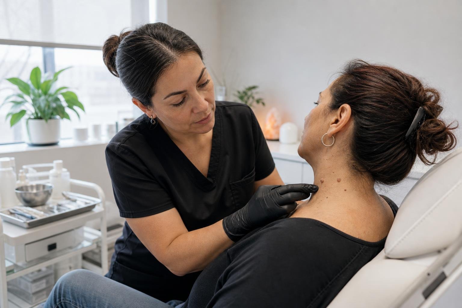
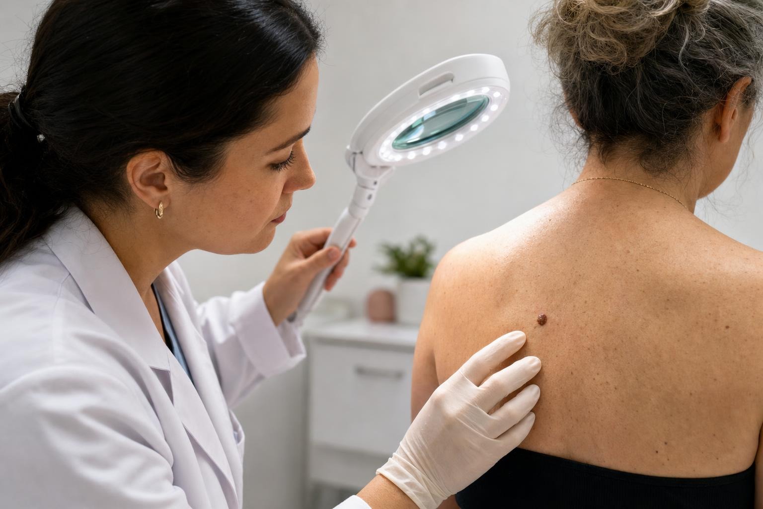
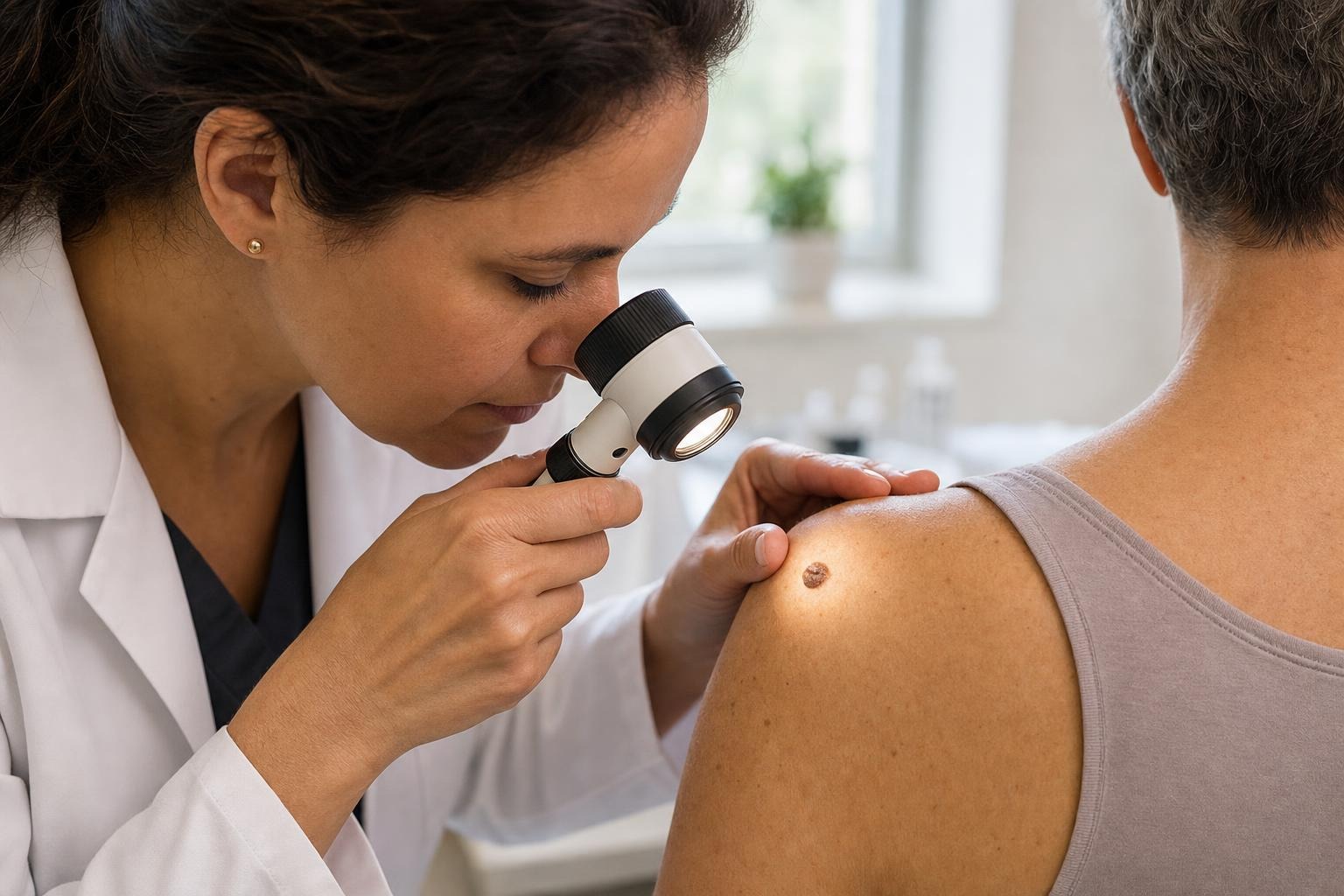
Clinicians assess more than colour and shape. They consider when the spot appeared, how it has changed, whether it causes symptoms and how it compares with the rest of the person’s skin. A physician or dermatologist may use magnification called dermoscopy. If uncertainty remains, a biopsy may be required to identify the lesion accurately.
An online photograph or visual checklist can help someone decide to seek care, but it cannot reliably exclude cancer. This is especially important before any removal procedure: destroying a suspicious spot without an appropriate assessment could delay diagnosis and eliminate tissue that might otherwise be examined.
Changes that deserve prompt medical assessment
The ABCDE guide is a useful framework for examining pigmented spots. The American Academy of Dermatology explains these melanoma warning signs:
- A — Asymmetry: One half does not resemble the other.
- B — Border: The edge is irregular, blurred, scalloped or poorly defined.
- C — Colour: Several colours appear within one lesion, or its colour changes.
- D — Diameter: Larger spots may warrant attention, although melanoma can be smaller than 6 millimetres.
- E — Evolving: The growth changes in size, shape, colour, elevation, surface or symptoms.
Evolution is particularly important. A spot that has looked the same for years is generally less concerning than one that has changed noticeably over several weeks or months. Not every change means cancer, but change creates a reason to investigate rather than guess.
The “ugly duckling” sign offers another practical test. Most of a person’s moles tend to share a general pattern. A lesion that is noticeably darker, larger, more irregular or otherwise unlike the others may deserve assessment even if it does not satisfy every ABCDE criterion.
Other reasons to seek medical advice include:
- Bleeding without a clear injury or repeatedly after minor contact
- A sore, crust or ulcer that does not heal
- Rapid growth or a newly raised area within an existing spot
- Persistent itching, pain or tenderness
- Pigment spreading beyond the original border
- A new and unusual pigmented lesion in adulthood
Skin cancer is not always dark. Some melanomas are pink, red or close to skin colour, and non-melanoma skin cancers may appear as persistent scaly patches or sores. The Canadian Cancer Society’s melanoma information provides additional signs and symptoms to discuss with a healthcare professional.
What to do when you notice a spot in Hamilton
Begin by leaving the lesion intact. Do not cut it, tie it off, burn it or apply a corrosive product while its identity is uncertain. Irritating the area can cause bleeding and inflammation, making later assessment more difficult.
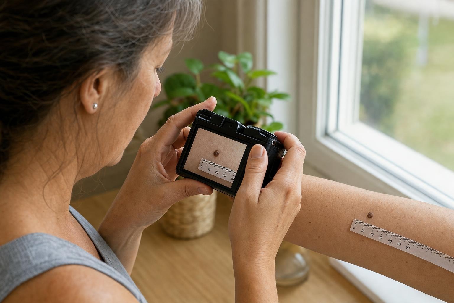
Next, document what you see. Take a clear photograph in even light, with a ruler nearby for scale, and record the date. Photograph the same angle under similar lighting if you check it again. This creates a more useful comparison than relying on memory. Avoid repeatedly handling or scraping the growth between photographs.
Consider four questions:
- Is it new, or has it been present and stable for years?
- Has its size, colour, border, thickness or surface changed?
- Does it stand out from your other spots?
- Is it bleeding, painful, persistently itchy or failing to heal?
If any answer raises concern—or if you simply cannot tell what the growth is—contact a physician or another qualified medical professional. Mention the change and timing when requesting an appointment rather than describing the issue only as cosmetic. A clear history helps the clinician decide how quickly it should be reviewed.
People with many moles, a personal or family history of melanoma, substantial ultraviolet exposure or immune suppression may need individualized advice about skin surveillance. A healthcare professional can recommend an appropriate schedule based on personal risk rather than a one-size-fits-all rule.
Where professional removal fits
A confirmed benign growth does not always need treatment. Removal is usually considered when it catches on clothing, becomes repeatedly irritated or causes cosmetic concern. The trade-offs should still be discussed: any skin procedure may result in temporary tenderness, a visible mark, pigment change or scarring.
Vicaria Health provides professional removal of skin tags, milia, ruby points and seborrheic keratoses in Hamilton, with services listed from $20 per lesion and no referral required. Same-day removal may be available when a lesion is suitable. Details about the local service are available through Vicaria Health.
That service distinction is important. Cosmetic removal is not a substitute for medical evaluation of an uncertain or suspicious lesion. If a spot has concerning features, has changed or might be a mole rather than a known benign keratosis, medical assessment should come first. Once the growth has been identified as benign, a removal conversation can focus appropriately on irritation, appearance, expected healing and possible skin changes.
Frequently asked questions
Can a seborrheic keratosis become melanoma?
A seborrheic keratosis is benign and does not turn into melanoma. The concern is misidentification: melanoma or another skin cancer may resemble a keratosis, and a cancerous lesion can occasionally occur near a benign growth. A new, changing or atypical spot should therefore be assessed rather than assumed to be harmless.
Can you identify the difference by touch?
Not reliably. A rough, waxy surface favours a keratosis, while many moles feel smooth, but there are exceptions. Some keratoses are thin and flat, and some benign moles are raised or textured. Touch cannot reveal the type of cells involved or rule out cancer.
Does a dark colour automatically mean a lesion is dangerous?
No. Benign keratoses and moles can be very dark, particularly on deeper skin tones. Conversely, melanoma can be pale, pink or red. Uneven colour, recent darkening and a spot that differs from the surrounding pattern are more informative than darkness alone.
Is bleeding always a sign of skin cancer?
A benign raised growth may bleed after being scratched or caught on clothing. Unexplained bleeding, repeated bleeding after minimal contact, or a sore that does not heal is more concerning. Arrange an assessment instead of repeatedly covering or treating the area without knowing the cause.
Can I remove a suspected keratosis at home?
Home cutting, tying, freezing or acid-based treatments can cause burns, infection, bleeding and scarring. More importantly, self-treatment may delay assessment of a lesion that is not actually a keratosis. The U.S. Food and Drug Administration warns against products marketed for removing moles and other skin lesions. Keep an uncertain growth intact and seek professional guidance.
Do all benign keratoses and moles need removal?
No. Stable, symptom-free benign growths can usually be left alone. Removal may make sense when a confirmed benign lesion is repeatedly irritated or unwanted for cosmetic reasons. The appropriate next step depends first on confidence about what the lesion is, then on personal preference and the expected procedural trade-offs.