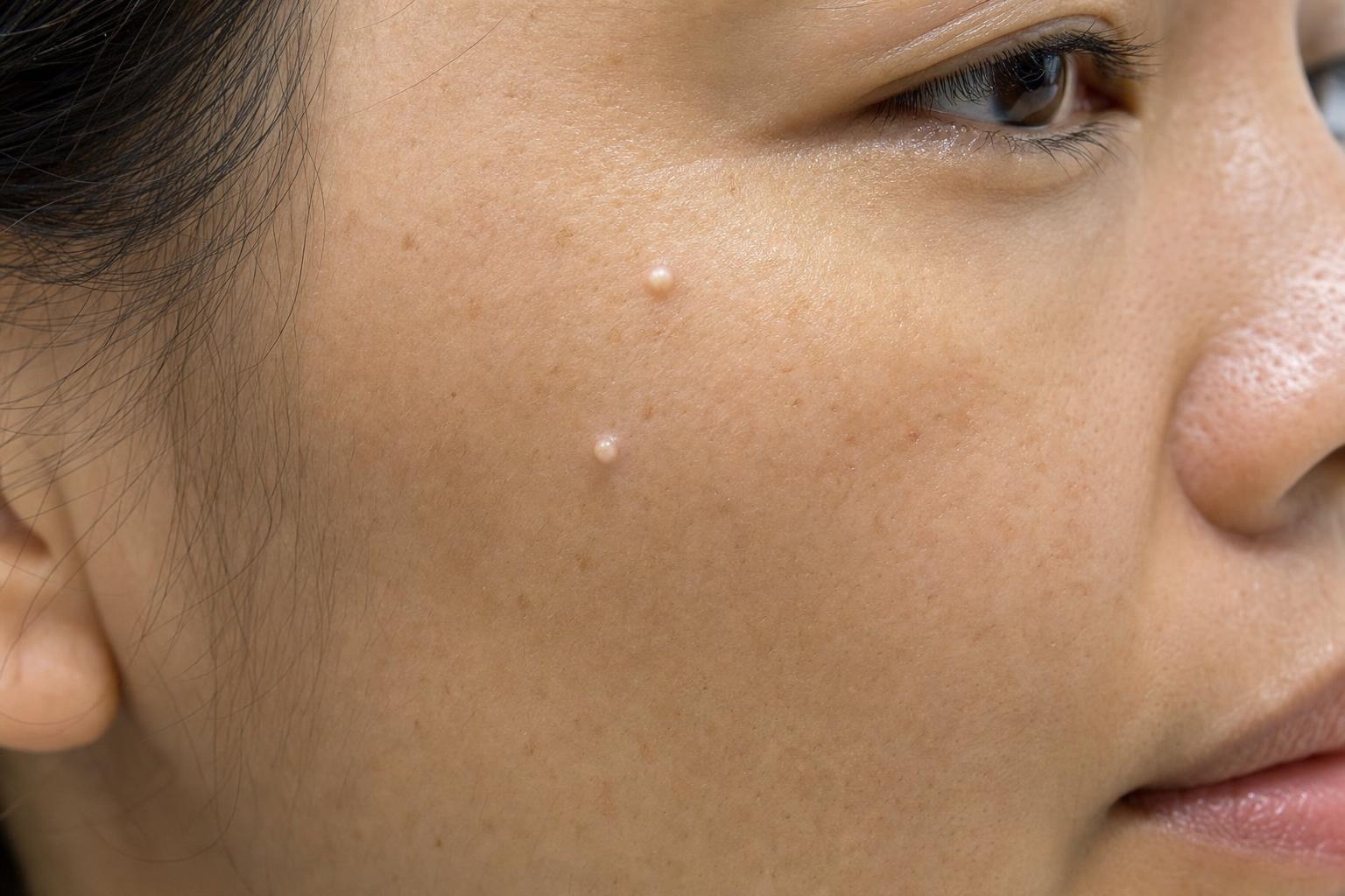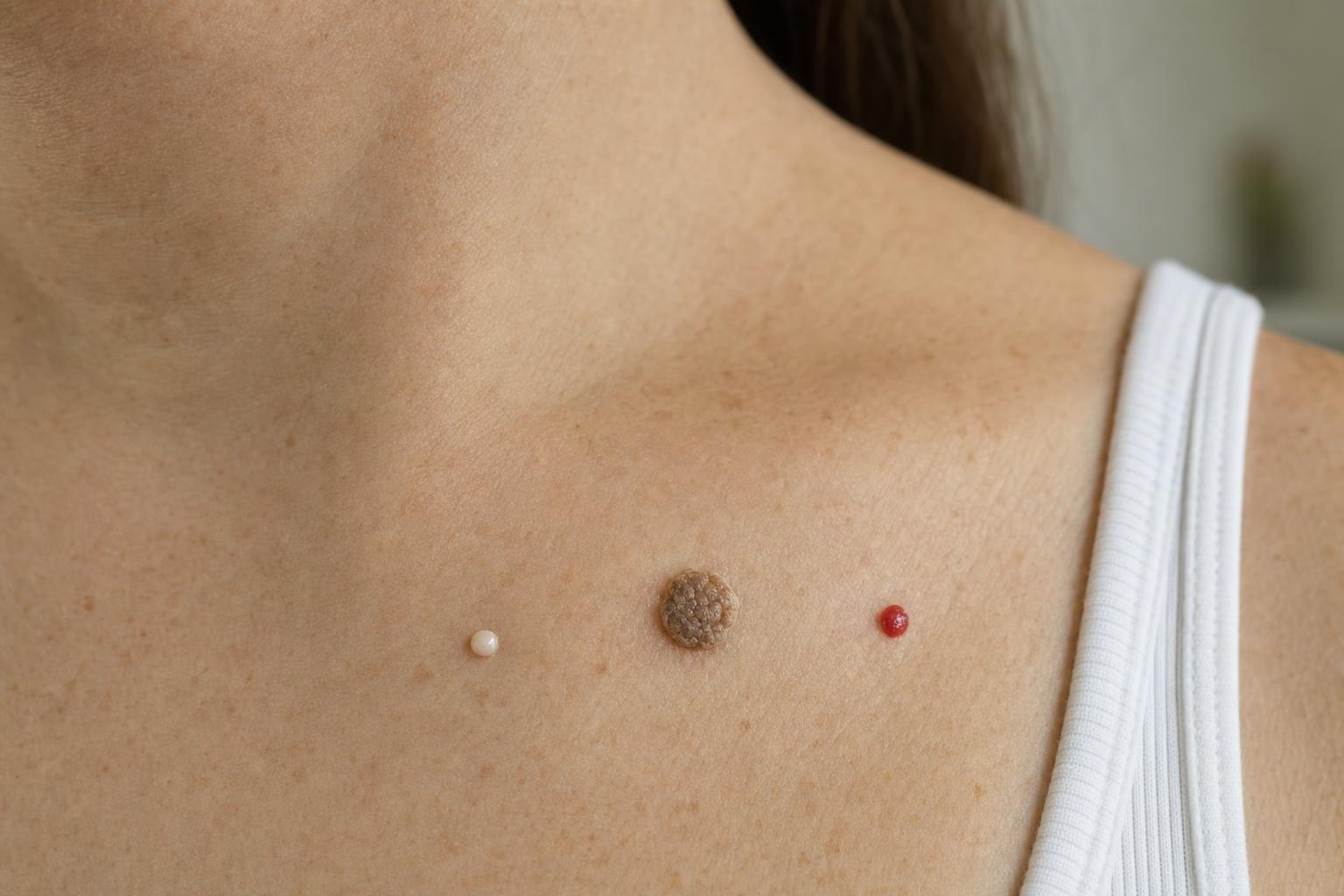
Milia are typically tiny white keratin cysts, seborrheic keratoses are waxy or rough growths that can look “stuck on,” and ruby points are small red vascular spots commonly known as cherry angiomas. Although these features can help you describe a lesion, appearance alone cannot confirm what it is. A new, changing, bleeding, or unusual growth should be assessed before cosmetic removal.
That distinction matters because several unrelated skin conditions can resemble one another. A dark seborrheic keratosis may be mistaken for a mole, a ruby point can bleed after being scratched, and milia near the eyes are sometimes confused with acne. Knowing the usual patterns helps you ask better questions without encouraging risky self-diagnosis.
Key takeaways
- Milia are firm, white or yellowish bumps containing keratin rather than acne fluid.
- Seborrheic keratoses are usually tan, brown, or dark growths with a waxy, scaly, or pasted-on surface.
- Ruby points are red to purple vascular spots, often called cherry angiomas.
- Sudden change, irregular colour, persistent bleeding, ulceration, or uncertainty warrants medical assessment.
- Cutting, burning, freezing, squeezing, or tying off an unidentified lesion at home can cause injury and delay diagnosis.
How the three lesions usually differ
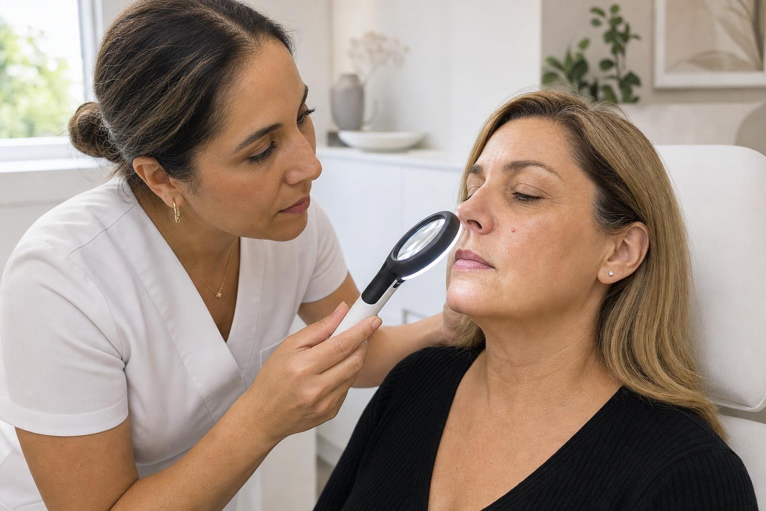
The most useful initial clues are colour, surface, firmness, location, and how the spot has behaved over time. None is decisive by itself, but the combination can narrow the possibilities.
Milia: small, firm keratin cysts
A milium is a small cyst containing keratin, the protein found in the outer layer of skin. Milia generally appear as pearly white, cream, or pale yellow bumps. They are often only a few millimetres wide and feel firm rather than soft or fluid-filled.
They commonly occur around the eyelids, cheeks, forehead, and nose. Unlike whiteheads, primary milia do not form because a pore is clogged with oil and bacteria. They usually have no visible opening and cannot be emptied like a typical pimple. Squeezing often produces redness or broken skin without removing the cyst.
Milia can occur at any age. Some develop spontaneously, while others appear after skin injury or inflammation. The DermNet overview of milia explains the different forms and their typical clinical appearance.
Seborrheic keratoses: waxy or rough growths
A seborrheic keratosis is a common benign growth that becomes more frequent with age. It may be skin-coloured, tan, brown, grey, or nearly black. Its surface can be smooth and waxy, rough, crumbly, or wart-like. Many look as though they have been pasted or stuck onto the skin.
These growths often appear on the trunk, scalp, face, neck, or shoulders. They can be flat at first and become thicker over time. A person may have one growth or many, and the lesions do not necessarily look identical.
Friction from a collar, bra strap, or waistband can irritate a raised growth. Irritation does not automatically make it dangerous, but a dark or inflamed lesion can be difficult to distinguish from other conditions. DermNet provides clinical examples in its seborrheic keratosis resource.
Ruby points: small vascular spots
“Ruby point” is a descriptive term often used for a cherry angioma. This is a benign cluster of small blood vessels near the skin’s surface. The spots are typically bright red when small, although larger or older ones may look deep red, burgundy, or purple. They can be flat or slightly raised and commonly occur on the torso, arms, shoulders, and neck.
Cherry angiomas tend to become more numerous with age. Because they contain blood vessels, they may bleed if scratched, shaved over, or caught on clothing. Recurrent trauma can make an otherwise harmless spot inconvenient.
Colour is the clearest contrast with milia, which are pale and solid, and most seborrheic keratoses, which have a visibly textured surface. The DermNet guide to cherry angiomas offers additional clinical context.
Why visual identification has limits
Online photographs can show common patterns, but lighting, skin tone, inflammation, image quality, and lesion age all affect appearance. A very small seborrheic keratosis may initially look smooth. Dried blood can make a vascular spot appear black. Milia can also resemble other small cysts or benign bumps.
The history of a lesion is often as important as its appearance. A professional may ask when it appeared, whether it has grown, whether the colour or border changed, and whether it hurts, itches, crusts, or bleeds without being disturbed. They may also consider medications, previous skin conditions, sun exposure, and personal or family history.
A cosmetic label should never be assumed when a lesion is new or atypical. Even people who have had benign growths before can develop an unrelated condition later. When the diagnosis is uncertain, assessment takes priority over removal.
When a lesion needs medical attention
Arrange medical assessment if a spot is changing noticeably, has several colours, develops an irregular border, becomes persistently painful, forms an open sore, or bleeds without scratching or friction. Rapid growth also deserves attention. These signs do not prove skin cancer, but they should not be dismissed as cosmetic.
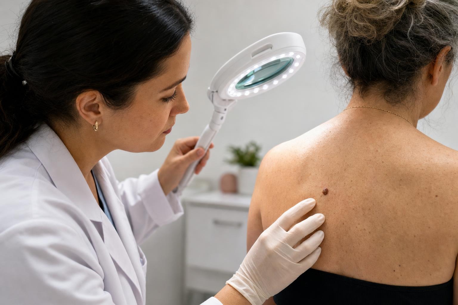
A dark growth that looks different from a person’s other spots—the so-called “ugly duckling”—is another reason to seek care. For pigmented lesions, the American Academy of Dermatology explains the ABCDE warning signs of melanoma, including asymmetry, border irregularity, colour variation, diameter, and evolution.
Location can change the level of concern or the type of care required. Growths on an eyelid margin, very close to the eye, inside the nose or mouth, or in another sensitive area may need evaluation by an appropriately qualified medical professional. The same applies if a person has a bleeding disorder, takes medication that affects bleeding, has impaired healing, or is immunocompromised.
If you are uncertain whether a spot is a long-standing benign lesion or something new, do not try to remove it first. Trauma can alter its appearance and make subsequent assessment more difficult.
How professional assessment and removal work
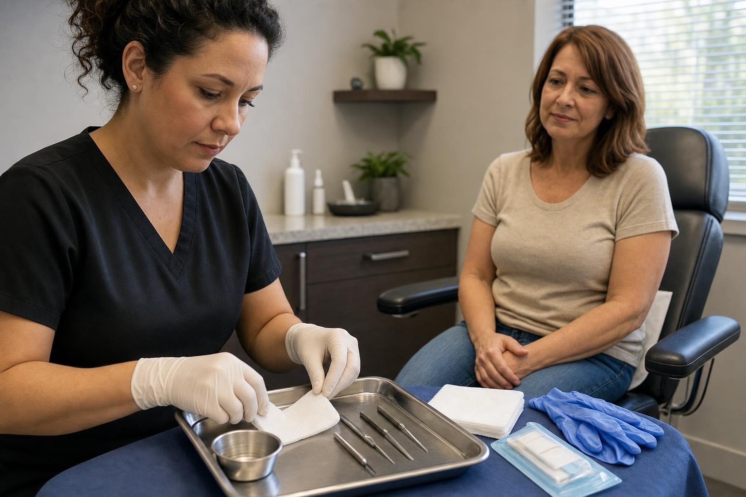
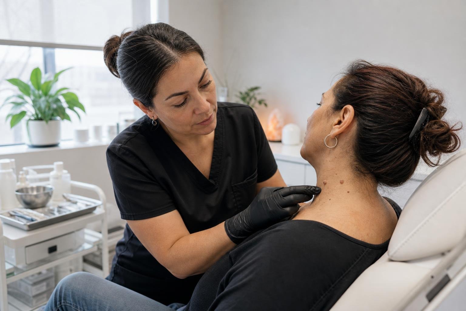
An appointment should begin with identifying whether the lesion has the typical features of a benign growth and whether removal is appropriate. The practitioner may inspect it under bright light, ask about changes, and determine whether referral for medical evaluation is the safer next step.
Removal is not always necessary. A stable lesion that causes no discomfort can often be left alone. People commonly consider removal when a confirmed benign growth catches on clothing, becomes irritated during shaving, sits in a cosmetically sensitive location, or causes self-consciousness.
The appropriate technique depends on the lesion type, depth, size, location, and number of growths. Milia are generally opened and extracted by a trained professional rather than squeezed through intact skin. Seborrheic keratoses may be treated using methods that remove or destroy the raised tissue. Vascular lesions require an approach suitable for blood-vessel tissue. No single method is right for every spot, and treatment around the eyes or on highly visible areas calls for particular care.
Before proceeding, ask what result is realistic and what aftercare will involve. Temporary redness, tenderness, crusting, or colour change can occur after many skin procedures. Any break in the skin also carries some risk of infection or scarring. Individual healing varies with skin type, lesion location, treatment method, and aftercare.
Vicaria Health provides professional removal of skin tags, milia, ruby points, and seborrheic keratoses in Hamilton without requiring a referral. Pricing starts from $20 per lesion, but suitability and the scope of treatment depend on assessment. Information about the practice and its services is available through Vicaria Health.
What not to do at home
Do not cut a suspected skin lesion with scissors or a blade. It is difficult to control bleeding, maintain sterile conditions, or know how deeply the tissue extends. This is especially dangerous for a vascular spot, which may bleed more than expected.
Avoid tying off, puncturing, burning, or applying acids to an unidentified growth. Products marketed for warts or skin tags are not automatically suitable for milia, seborrheic keratoses, or vascular lesions. Applying them to the wrong condition can cause a chemical burn, pigment change, infection, or scarring.
Freezing kits also present problems. Their effect can be difficult to limit to the lesion, and partial treatment may leave an irritated or altered growth behind. Never use these products near the eyes unless specifically directed by an appropriate healthcare professional.
Milia should not be treated like pimples. Forceful squeezing can damage the surrounding skin without creating an opening through which the keratin can escape. Gentle skincare may reduce irritation, but it cannot instantly extract an established cyst.
If a spot is repeatedly caught by clothing, protect it temporarily with a clean, non-stick dressing and arrange an assessment. If it bleeds after accidental trauma, apply steady pressure with clean gauze. Seek medical care if bleeding is heavy, does not stop, or recurs without an obvious cause.
Preparing for an appointment in Hamilton
Before an assessment, note how long the lesion has been present and whether its size, colour, texture, or symptoms have changed. A dated photograph can be useful when change has occurred gradually, provided the image is not treated as a substitute for examination.
Bring a list of relevant medications and disclose conditions that affect bleeding, immune function, skin healing, or scarring. Mention previous reactions to local treatments or adhesives. Avoid deliberately irritating the lesion before the visit, and arrive without concealer or heavy skincare over the area when practical.
Useful questions include:
- Does this lesion have the usual features of a benign condition?
- Is removal appropriate, or should it be medically assessed first?
- What should I expect while the area heals?
- How should I protect the treated site from friction or sun exposure?
- Which changes after treatment would require follow-up?
A careful consultation should leave room for referral rather than treatment when the lesion is unclear. That is an important safety decision, not an inconvenience.
Frequently asked questions
Are milia a form of acne?
No. Milia are small cysts filled with keratin and do not arise from the same inflammatory process as acne. They can occur alongside acne, but squeezing them like whiteheads commonly injures the surrounding skin without removing the cyst.
Can a seborrheic keratosis turn into cancer?
Seborrheic keratoses are benign growths. The practical concern is that another condition can resemble one, or a suspicious lesion can appear near an existing keratosis. A growth that changes, looks unusual, or cannot be confidently identified should be medically assessed.
Why does a ruby point bleed so easily?
A cherry angioma consists of small blood vessels close to the skin’s surface. Scratching, shaving, or friction can break the surface and cause bleeding. Bleeding after obvious trauma can occur, but spontaneous or repeated bleeding should be evaluated.
Will these lesions disappear without treatment?
Some milia resolve on their own, particularly in infants, but others persist. Seborrheic keratoses and cherry angiomas generally remain unless treated. Leaving a stable, confirmed benign lesion alone is often reasonable if it is not irritated or bothersome.
Can several lesions be removed during one visit?
Potentially, but this depends on their type, location, number, and whether each one is suitable for removal. Similar-looking spots should still be considered individually. An assessment can establish which lesions may be treated and whether any require medical referral.
Does removal prevent new lesions from forming?
Removal addresses the treated lesion; it does not necessarily prevent new milia, seborrheic keratoses, or cherry angiomas from developing elsewhere. Some people are prone to developing multiple benign growths over time. Continue monitoring the skin and have new or changing spots assessed rather than assuming they match previous ones.